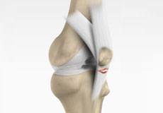
Patella tendon rupture is the rupture of the tendon that connects the patella (knee cap) to the top portion of the tibia (shin bone). The patellar tendon works together with the quadriceps muscle and the quadriceps tendon to allow your knee to straighten out.
Patellar tendon tear most commonly occurs in middle-aged people who participate in sports which involve jumping and running. Patellar tendon can be ruptured by several reasons such as by fall, direct blow to the knee, or landing on the foot awkwardly from a jump. Other causes include patellar tendonitis (inflammation of patellar tendon), diseases such as rheumatoid arthritis, diabetes mellitus, infection, and chronic renal failure. Use of medications such as steroids can cause increased muscle and tendon weakness.
When the patellar tendon tears, the patella may lose its anchoring support to the tibia as a result when the quadriceps muscle contracts the patella may move up into the thigh. You are unable to straighten your knee and upon standing the knee buckles upon itself. In addition to this you may have pain, swelling, tenderness, a tearing or popping sensation, bruising, and cramping.
Patellar tendon tear can be a partial or a complete tear. In partial tear, some of the fibres in the tendon are torn, but the soft tissue is not damaged. In complete tear, the soft tissues are disrupted into two pieces.
To identify a patellar tendon tear your doctor will ask about your medical history and perform a physical examination of your knee. Some imaging tests, such as an X-ray or magnetic resonance imaging (MRI) scan may be ordered to confirm the diagnosis. X-ray of the knee is taken to know the position of the kneecap and MRI scan to know the extent and location of the tear.
Patellar tendon rupture can be treated by non-surgical and surgical methods. Non-surgical treatment involves use of braces or splints to immobilise the knee. Physical therapy may be recommended to restore the strength and increase range of motion of the knee.
Surgery is performed on an outpatient basis and not arthroscopically since the tendon is present outside the joint. The goal of the surgery is to reattach the torn tendon to knee cap and to restore the normal function in the affected leg. The procedure is performed under regional or general anaesthesia and an incision is made on the front of the knee to expose the tendon rupture. Holes are made in the patella and strong sutures are tied to the tendon and then threaded through these holes. These sutures are tied in place to pull the torn edge of the tendon back to its normal position on the kneecap.
Severe damage can make the patellar tendon very short, and in such cases reattachment will be difficult. Your surgeon may attach a tissue taken from a donor (allograft) to lengthen the tendon.
Complications after the repair include weakness and loss of motion. In some cases, the tendon which re-attached may detach from the knee cap or re-tears may also occur. Other complications such as infection and blood clot may be observed.
Following surgery, a brace may be needed to protect the healing tendon. Complete healing of the tendon will take about 6 months.


